
Berkas:Coronavirus. SARS-CoV-2.png

Ukuran asli (2.048 × 2.048 piksel, ukuran berkas: 4,54 MB, tipe MIME: image/png)
Ringkasan
| Deskripsi |
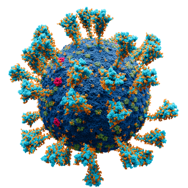
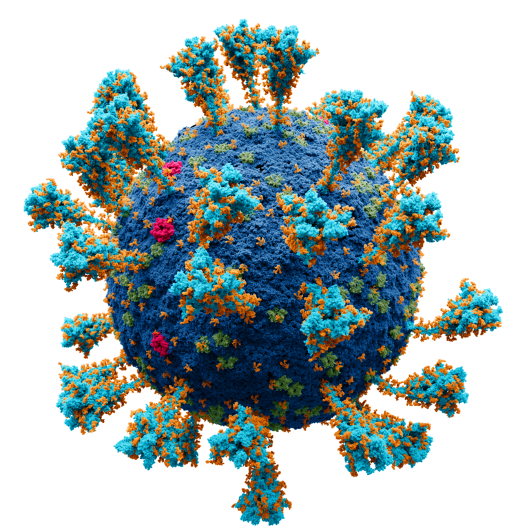
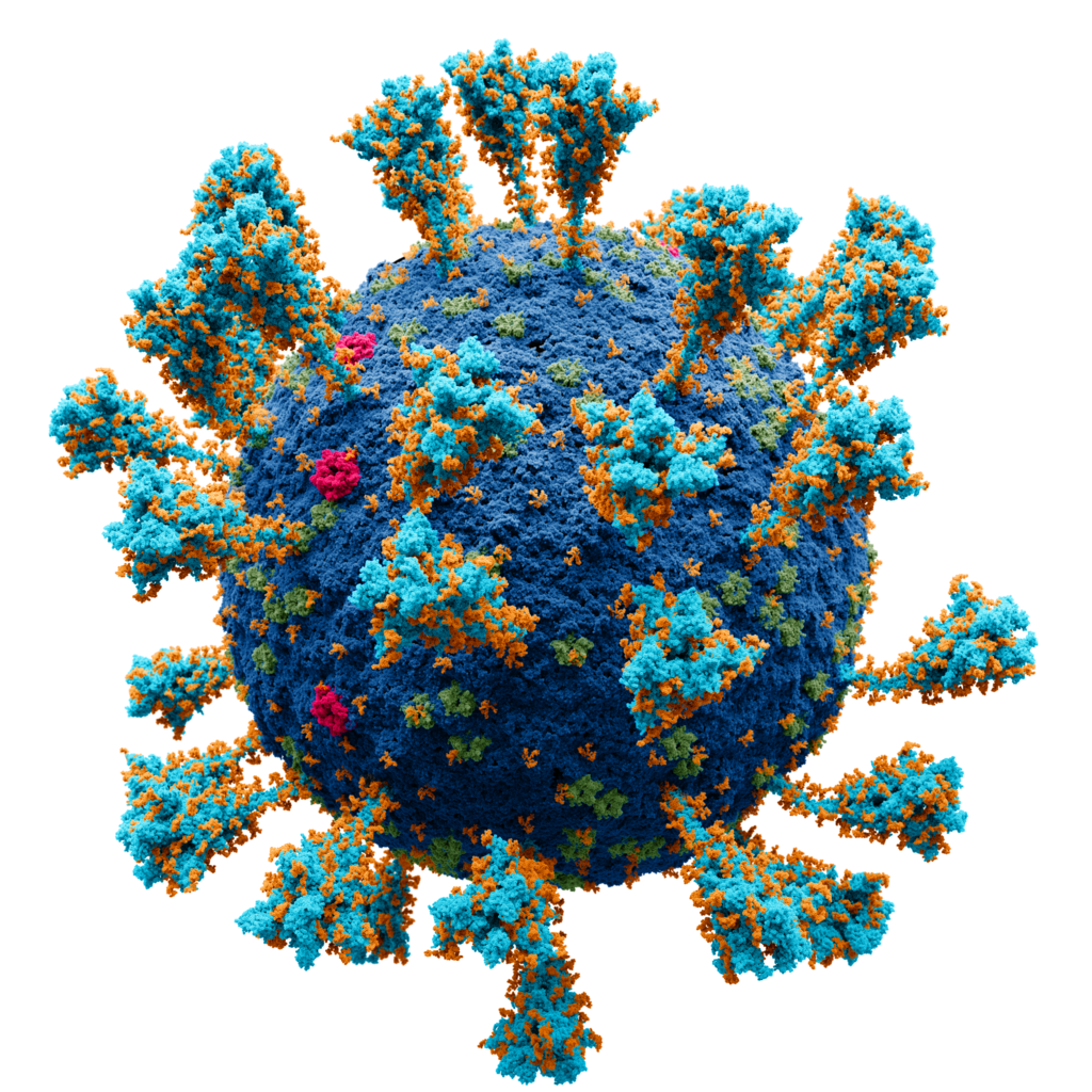
Deutsch: Wissenschaftlich genaues Atommodell der äußeren Struktur des SARS Coronavirus 2 (SARS-CoV-2), einem Stamm (genetische Variante) des Coronavirus, der die Coronavirus-Krankheit (COVID-19) verursachte und erstmals im Dezember 2019 in Wuhan, China, identifiziert wurde.
Jeder einzelne Ort (amorpher Fleck) ist ein Atom von: kobalt: Virushülle
purpurrot: Hüllproteine
grün: Matrixproteine
türkis: Spike-Proteine English: Scientifically accurate atomic model of the external structure of the Severe Acute Respiratory Syndrome CoronaVirus 2 (SARS-CoV-2), a strain (genetic variant) of the coronavirus that caused coronavirus disease (COVID-19), first identified in Wuhan, China, during December 2019
Each separate locus (amorphous blob) is a molecule of: cobalt: membrane
crimson: E protein
green: M protein
turquoise : S (spike) glycoprotein Español: Modelo atómico de la estructura externa del SARS-CoV-2. Cada "bola" es un átomo.
Русский: Научно достоверная атомарная модель внешней структуры коронавируса (SARS-CoV-2). Каждый "шарик" — атом. Опубликовано на N+1.
Научный консультант:
Protein Data Bank: 2mls, 6y3y, 5x29, 6yyt Charmm-gui: 6vsb_1_1_1_S309
кобальт — мембрана.
бирюза — S-белок.
малиновый — E-белок.
зелёный — M-белок.
оранжевый — гликаны.
Проект был сделан в соответствии с научными источниками:
Первичные источники:
|
| Tanggal | |
| Sumber |
Karya sendiri. Scientific consultants:
|
| Pembuat | Alexey Solodovnikov (Idea, Producer, CG, Editor), Valeria Arkhipova (Scientific Сonsultant) |
| Izin (Menggunakan kembali berkas ini) |
Published by N+1 a popular science online publication of Russia (https://nplus1.ru/), protein models are derivative works of the free license site (freely available for both non-commercial and commercial use) |
| Versi lainnya |
|
Sources
Primary sources:
- https://www.ncbi.nlm.nih.gov/pmc/articles/PMC7489918/
- https://www.ncbi.nlm.nih.gov/pmc/articles/PMC7098027/
- https://www.ncbi.nlm.nih.gov/pmc/articles/PMC7605623/
The following structures from open sources were used in the work, Protein Data Bank (https://www.rcsb.org):
- 2mls (membrane bilayer complex with matrix metalloproteinase-12 at its beta-face) Koppisetti, R.K., Fulcher, Y.G., Prior, S.H., Lenoir, M., Overduin, M., Van Doren, S.R. 2014
- 6y3y (human coronavirus HKU1 haemagglutinin-esterase) Hurdiss, D.L., Drulyte, I., Pronker, M.F. 2020
- 5x29 (NMR structure of the SARS Coronavirus E protein pentameric ion channel) Torres, J., Surya, W., Li, Y. 2017
- 6yyt (Structure of replicating SARS-CoV-2 polymerase) Hillen, H.S., Kokic, G., Farnung, L., Dienemann, C., Tegunov, D., Cramer, P. 2020
- Charmm-gui: 6vsb_1_1_1_S309
Additional sources:
- Surya, W., Li, Y., Torres, J. Structural model of the SARS coronavirus E channel in LMPG micelles // Biochim. Biophys. Acta - Biomembr. – 2018. – Vol. 1860. – N. 6. – P. 1309–1317.
- Koppisetti, R. K., Fulcher, Y. G., Jurkevich, A., Prior, S. H., Xu, J., Lenoir, M., Overduin, M., Van Doren, S. R. Ambidextrous binding of cell and membrane bilayers by soluble matrix metalloproteinase-12 // Nat. Commun. – 2014. – Vol. 5. – P. 1–14.
- Hillen, H. S., Kokic, G., Farnung, L., Dienemann, C., Tegunov, D., Cramer, P. Structure of replicating SARS-CoV-2 polymerase // Nature. – 2020. – Vol. 584. – N. 7819. – P. 154–156.
- Harris, L. J., Larson, S. B., Hasel, K. W., McPherson, A. Refined structure of an intact IgG2a monoclonal antibody // Biochemistry. – 1997. – Vol. 36. – N. 7. – P. 1581–1597.
- Noreng, S., Bharadwaj, A., Posert, R., Yoshioka, C., Baconguis, I. Structure of the human epithelial sodium channel by cryo-electron microscopy // Elife. – 2018. – Vol. 7. – P. 1–23.
- Almond, A., DeAngelis, P. L., Blundell, C. D. Hyaluronan: The Local Solution Conformation Determined by NMR and Computer Modeling is Close to a Contracted Left-handed 4-Fold Helix // J. Mol. Biol. – 2006. – Vol. 358. – N. 5. – P. 1256–1269.
- Hurdiss, D. L., Drulyte, I., Lang, Y., Shamorkina, T. M., Pronker, M. F., van Kuppeveld, F. J. M., Snijder, J., de Groot, R. J. Cryo-EM structure of coronavirus-HKU1 haemagglutinin esterase reveals architectural changes arising from prolonged circulation in humans // Nat. Commun. – 2020. – Vol. 11. – N. 1. – P. 1–10.
- Yan, Renhong, Yuanyuan Zhang, Yaning Li, Lu Xia, Yingying Guo, Q. Z. Structural basis for the recognition of SARS-CoV-2 by full-length human ACE2 // Science (80-. ). – 2020. – Vol. 3. – N. 3. – P. 1–8.
- Javitt, G., Khmelnitsky, L., Albert, L., Bigman, L. S., Elad, N., Morgenstern, D., Ilani, T., Levy, Y., Diskin, R., Fass, D. Assembly Mechanism of Mucin and von Willebrand Factor Polymers // Cell. – 2020. – Vol. 183. – N. 3. – P. 717-729.e16.
- Daniel Wrapp, Nianshuang Wang, Kizzmekia S. Corbett, Jory A. Goldsmith, Ching-Lin Hsieh, Olubukola Abiona, B. S. G., McLellan, and J. S. Cryo-EM structure of the 2019-nCoV spike in the prefusion conformation // Science (80-. ). – 2020. – Vol. 21. – N. 1. – P. 1–9.
- Wang, M. Y., Zhao, R., Gao, L. J., Gao, X. F., Wang, D. P., Cao, J. M. SARS-CoV-2: Structure, Biology, and Structure-Based Therapeutics Development // Front. Cell. Infect. Microbiol. – 2020. – Vol. 10. – N. November. – P. 1–17. (https://pubmed.ncbi.nlm.nih.gov/33324574/)
- Yao, H., Song, Y., Chen, Y., Wu, N., Xu, J., Sun, C., Zhang, J., Weng, T., Zhang, Z., Wu, Z., Cheng, L., Shi, D., Lu, X., Lei, J., Crispin, M., Shi, Y., Li, L., Li, S. Molecular Architecture of the SARS-CoV-2 Virus // Cell. – 2020. – Vol. 183. – N. 3. – P. 730-738.e13.
- Oostra, M., de Haan, C. A. M., de Groot, R. J., Rottier, P. J. M. Glycosylation of the Severe Acute Respiratory Syndrome Coronavirus Triple-Spanning Membrane Proteins 3a and M // J. Virol. – 2006. – Vol. 80. – N. 5. – P. 2326–2336. (https://europepmc.org/article/MED/16474139)
- B.W. Neuman, M. J. B. Supramolecular Architecture of the Coronavirus Particle // Adv. Virus Res. – 2020. – Vol. 96. – P. 1–27 (https://www.ncbi.nlm.nih.gov/pmc/articles/PMC7112365/, https://europepmc.org/article/PMC/1563832)
- Neuman, B. W., Kiss, G., Kunding, A. H., Bhella, D., Baksh, M. F., Connelly, S., Droese, B., Klaus, J. P., Makino, S., Sawicki, S. G., Siddell, S. G., Stamou, D. G., Wilson, I. A., Kuhn, P., Buchmeier, M. J. A structural analysis of M protein in coronavirus assembly and morphology // J. Struct. Biol. – 2011. – Vol. 174. – N. 1. – P. 11–22. (https://www.ncbi.nlm.nih.gov/pmc/articles/PMC4486061/)
- Yu, A., Pak, A. J., He, P., Monje-Galvan, V., Casalino, L., Gaieb, Z., Dommer, A. C., Amaro, R. E., Voth, G. A. A multiscale coarse-grained model of the SARS-CoV-2 virion // Biophys. J. – 2021. – Vol. 120. – N. 6. – P. 1097–1104 (https://europepmc.org/article/PMC/PMC7695975, https://search.bvsalud.org/global-literature-on-novel-coronavirus-2019-ncov/resource/en/covidwho-947143)
- Yao, H., Song, Y., Chen, Y., Wu, N., Xu, J., Sun, C., Zhang, J., Weng, T., Zhang, Z., Wu, Z., Cheng, L., Shi, D., Lu, X., Lei, J., Crispin, M., Shi, Y., Li, L., Li, S. Molecular architecture of the SARS-CoV-2 virus // Cell. – 2020. – Vol. 183. – N. 3. – P. 730–738 (https://www.sciencedirect.com/science/article/pii/S0092867420311594)
- Choi, Y. K., Cao, Y., Frank, M., Woo, H., Park, S. J., Yeom, M. S., Croll, T. I., Seok, C., Im, W. Structure, Dynamics, Receptor Binding, and Antibody Binding of the Fully Glycosylated Full-Length SARS-CoV-2 Spike Protein in a Viral Membrane // J. Chem. Theory Comput. – 2021. – Vol. 17. – N. 4. – P. 2479–2487 (https://www.researchgate.net/publication/349986293_Structure_Dynamics_Receptor_Binding_and_Antibody_Binding_of_the_Fully_Glycosylated_Full-Length_SARS-CoV-2_Spike_Protein_in_a_Viral_Membrane)
Lisensi
- Anda diizinkan:
- untuk berbagi – untuk menyalin, mendistribusikan dan memindahkan karya ini
- untuk menggubah – untuk mengadaptasi karya ini
- Berdasarkan ketentuan berikut:
- atribusi – Anda harus mencantumkan atribusi yang sesuai, memberikan pranala ke lisensi, dan memberi tahu bila ada perubahan. Anda dapat melakukannya melalui cara yang Anda inginkan, namun tidak menyatakan bahwa pemberi lisensi mendukung Anda atau penggunaan Anda.
- berbagi serupa – Apabila Anda menggubah, mengubah, atau membuat turunan dari materi ini, Anda harus menyebarluaskan kontribusi Anda di bawah lisensi yang sama seperti lisensi pada materi asli.
Penilaian
|


Riwayat berkas
Klik pada tanggal/waktu untuk melihat berkas ini pada saat tersebut.
| Tanggal/Waktu | Miniatur | Dimensi | Papaké | Komentar | |
|---|---|---|---|---|---|
| terkini | 9 Januari 2022 22.17 | | 2.048 × 2.048 (4,54 MB) | Jul059 | Lossless file size reduction |
| 24 September 2021 03.58 |  | 2.048 × 2.048 (4,6 MB) | Iketsi | lossless compression | |
| 15 Juni 2021 16.06 |  | 2.048 × 2.048 (5,34 MB) | AlexeySolodovnikov | fix color bug | |
| 13 Juni 2021 14.28 |  | 2.048 × 2.048 (5,34 MB) | AlexeySolodovnikov | Мы обновили модель. В роли нашего научного консультанта выступил доктор биологических наук, специалист в области вирусологии, Никитин Н. А. и к.х.н специалист по молекулярному моделированию поверхностных вирусных белков Борисевич С.С. Под их руководством в модель были внесены следующие правки: Изменено количество S-белков с 90 до 38, количество M-белков было увеличено до 1000, а E-белков, как минорных компонентов мембраны, снижено до 15, HE-белок удалён. Также была принята во внимание шарни... | |
| 17 Méi 2021 11.06 |  | 2.048 × 2.048 (16,04 MB) | AlexeySolodovnikov | add alpha | |
| 4 Méi 2021 18.41 |  | 2.048 × 2.048 (16,04 MB) | AlexeySolodovnikov | Uploaded own work with UploadWizard |
Pranala
Halaman berikut menggunakan berkas ini:
Penggunaan berkas global
Wiki lain berikut menggunakan berkas ini:
- Penggunaan pada alt.wikipedia.org
- Penggunaan pada ar.wikipedia.org
- مراكز السيطرة على الأمراض والوقاية منها
- فيروس كورونا
- مستخدم:Amira Hashem1996/ملعب
- مناطق انتشار جائحة فيروس كورونا حسب الدولة والمنطقة
- عزل ووهان 2020
- قائمة حوادث كراهية الأجانب والعنصرية المرتبطة بجائحة فيروس كورونا
- مستشفى هوو شين شان
- مستشفى لي شين شان
- جائحة فيروس كورونا في العراق
- معهد ووهان لأبحاث الفيروسات
- جائحة فيروس كورونا في إيطاليا
- جائحة فيروس كورونا في الجزائر
- جائحة فيروس كورونا في اليونان
- اللجنة الوطنية للصحة (الصين)
- جائحة فيروس كورونا في الكويت
- جائحة فيروس كورونا في الكاميرون
- المركز الصيني لمكافحة الأمراض والوقاية منها
- جائحة فيروس كورونا في البوسنة والهرسك
- أثر جائحة فيروس كورونا على الحياة الاجتماعية
- مستشفى ووهان المركزي
- جائحة فيروس كورونا في الأردن
- أثر جائحة فيروس كورونا على الرياضة
- جائحة فيروس كورونا في السودان
- جائحة فيروس كورونا في فرنسا
- جائحة فيروس كورونا في إفريقيا
- جائحة فيروس كورونا في جمهورية الكونغو الديمقراطية
- جائحة فيروس كورونا في الغابون
- انهيار فندق شينجيا إكسبريس
- جائحة فيروس كورونا في توغو
- جائحة فيروس كورونا في غينيا
- جائحة فيروس كورونا في رواندا
- جائحة فيروس كورونا في ساحل العاج
- جائحة فيروس كورونا في ناميبيا
- جائحة فيروس كورونا في كينيا
- جائحة فيروس كورونا في مايوت
- جائحة فيروس كورونا في لا ريونيون
- قيود السفر بسبب جائحة فيروس كورونا
- جائحة فيروس كورونا في غينيا الاستوائية
- جائحة فيروس كورونا في جمهورية إفريقيا الوسطى
- جائحة فيروس كورونا في جمهورية الكونغو
- جائحة فيروس كورونا في سيشل
- جائحة فيروس كورونا في ليبيريا
- جائحة فيروس كورونا في الصومال
- جائحة فيروس كورونا في تنزانيا
- جائحة فيروس كورونا في كازاخستان
- جائحة فيروس كورونا في أوروبا
- لقاح كوفيد-19
- جائحة فيروس كورونا في أوقيانوسيا
- جائحة فيروس كورونا في كولومبيا
Lihat lebih banyak penggunaan global dari berkas ini.